Ameloblastoma is a rare, highly destructive jaw tumor. Dr. Manish Tiwari specializes in aggressively clearing the tumor to prevent recurrence, followed by immediate 3D microvascular bone reconstruction to perfectly restore your facial appearance.
A slowly enlarging, usually painless, hard lump on the lower jaw (mandible).
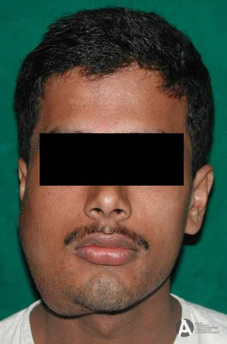
Noticeable distortion of the lower face as the tumor expands the bone outward.
Teeth in the affected area become loose or shift positions as bone is destroyed.
Often discovered by dentists incidentally as a "soap bubble" appearance on an OPG scan.
Though technically "benign" (non-cancerous), Ameloblastomas are locally aggressive. Simply scraping them out (enucleation) results in an unacceptably high recurrence rate. Definitive cure requires removing a segment of the jawbone.
Ameloblastoma cells infiltrate deep into the microscopic spaces of the jawbone far beyond what is visible on an X-ray. Inadequate, conservative surgery almost guarantees the tumor will return, causing even more damage.
The gold standard for solid or multicystic Ameloblastomas. Dr. Tiwari completely excises the tumor along with a 1cm to 1.5cm safety margin of visually healthy bone, guaranteeing the tumor is eradicated permanently.
Patients often fear that a resection means permanent facial deformity. By utilizing microvascular surgery, Dr. Tiwari instantly rebuilds the missing jawbone in the same sitting, so you wake up with your facial structure intact.
We utilize 3D Virtual Surgical Planning (VSP) to map the exact cuts and the precise microvascular reconstruction weeks before surgery.
Your CT scans are used to create a 3D computer model. We design custom 3D-printed cutting guides to ensure exact, symmetrical bone removal.
The diseased jaw segment containing the Ameloblastoma is completely removed, ensuring negative (clear) bone margins.
Healthy bone from your lower leg (fibula) is shaped with titanium plates to match your jaw's exact curve. Its blood vessels are connected to your neck via microsurgery, bringing the new bone to life.

Accurate imaging differentiates Ameloblastoma from simple dental cysts.
Our goal is complete return to normalcy, including your ability to chew solid food.