Expert surgical management of Odontogenic Keratocysts (OKC), Dentigerous Cysts, and other benign jaw lesions. Dr. Manish Tiwari utilizes advanced enucleation and bone-preserving techniques to eradicate cysts while protecting your teeth and facial nerves.
A slow-growing, hard bump on the upper or lower jawbone that gradually distorts the face.
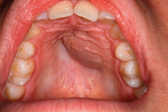
Teeth mysteriously shifting positions, becoming loose, or adult teeth failing to erupt.
Noticeable differences between the left and right sides of the jawline or cheek.
Numbness, tingling, or a "pins and needles" sensation in the lower lip or chin area.
While these cysts are benign (non-cancerous), they aggressively hollow out the jawbone. Our goal is to completely remove the cyst lining to prevent it from growing back, while saving the structural integrity of your jaw.
The primary treatment for most jaw cysts. The entire cyst is carefully separated from the bone cavity and shelled out intact. The surrounding bone is then meticulously scraped (curettage) to ensure no microscopic cyst cells remain.
When a cyst is so massive that removing it immediately might break the jaw or damage vital nerves, we use Marsupialization. We create a small opening to drain the cyst, relieving pressure so it shrinks over several months before final removal.
Highly aggressive cysts, like the Odontogenic Keratocyst (OKC), have "daughter cysts" that hide deep in the bone marrow. Dr. Tiwari uses a specialized bur to safely grind away a thin, 1-2mm layer of surrounding bone after enucleation.
Dr. Tiwari prioritizes an intraoral approach—performing the entire surgery from inside the mouth to ensure absolutely no visible scars on your face.
Before surgery, advanced 3D scans are used to map the exact location of the cyst and its proximity to tooth roots and the inferior alveolar nerve.
Operating entirely through the gums, the cyst is carefully separated from the bone and extracted. Impacted teeth causing the cyst (like wisdom teeth) are also removed.
Once the cyst is out, the empty void in the jaw is packed with advanced bone grafts and PRF (Platelet-Rich Fibrin) to stimulate the body to regenerate strong, new jawbone.
A lump in the jaw can be many things. Accurate diagnosis dictates the aggressiveness of the surgery.
Healing is usually fast, but protecting the weakened jaw is critical in the early stages.